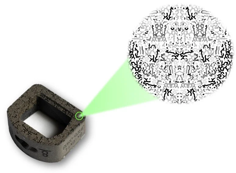
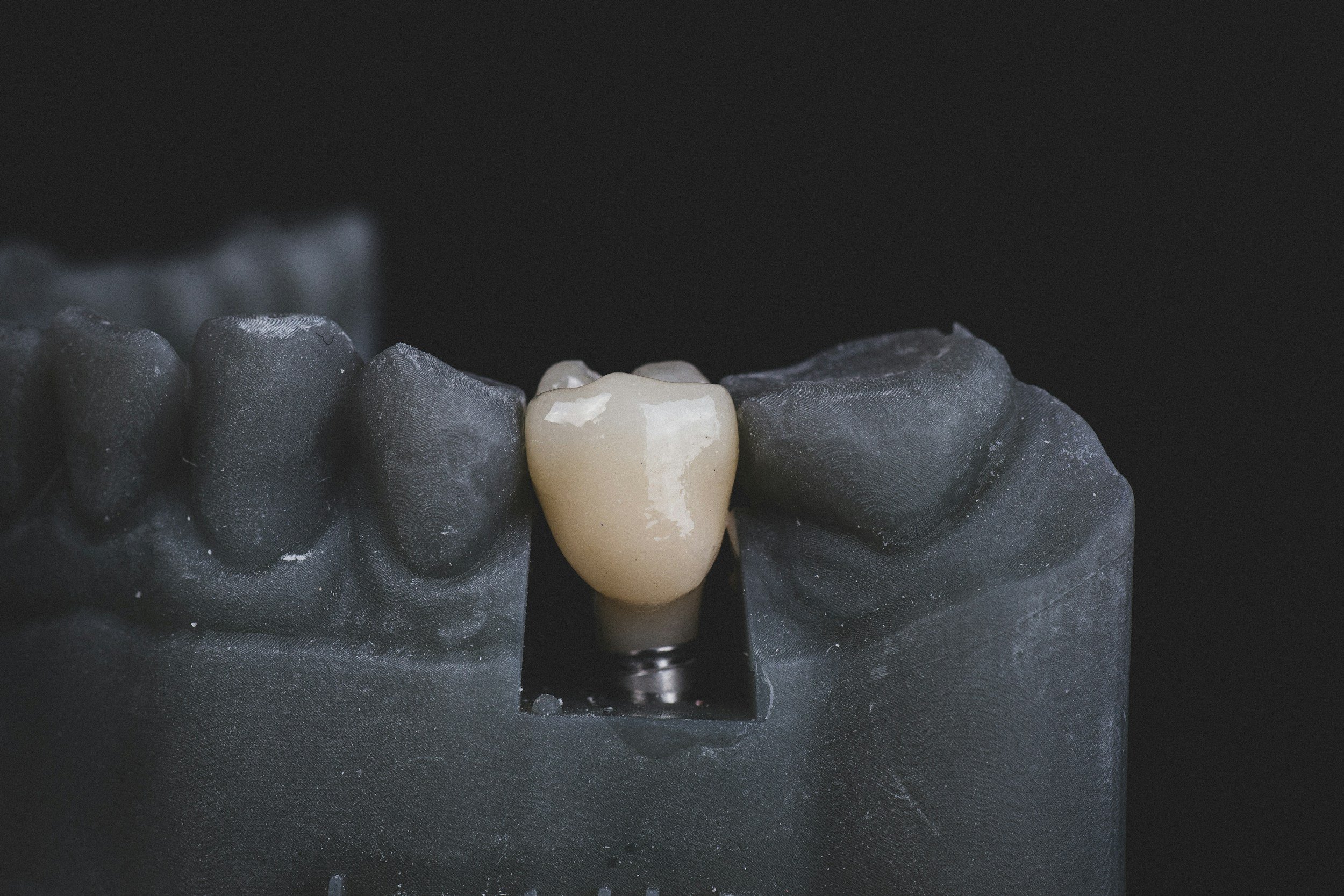
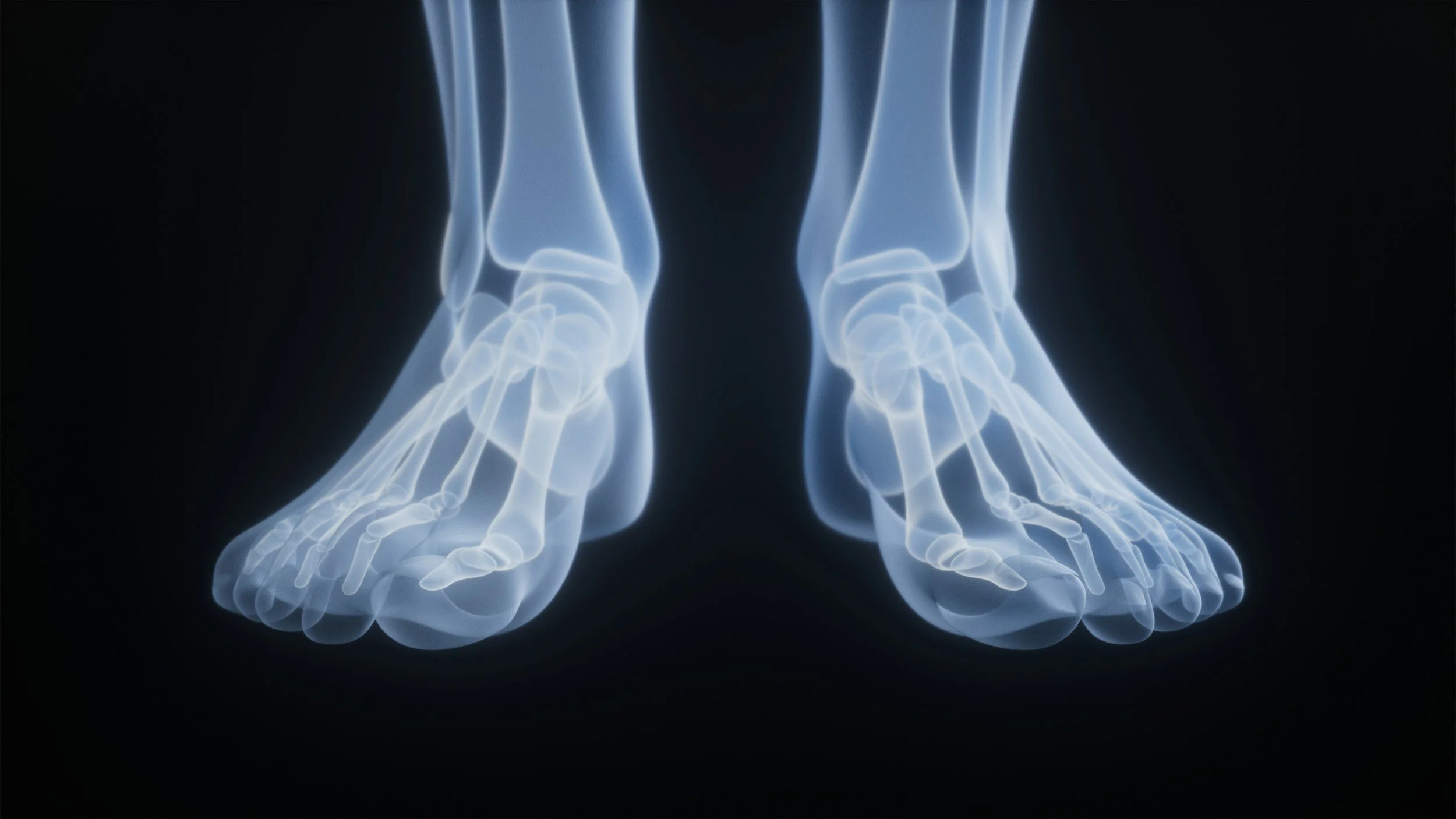
BioBraille™
BioBraille™ is the next generation of nanotechnology.
Macro-, micro-, and nanofeatures are “programmed” onto the implant surface utilizing a proprietary and advanced laser etching process. This approach is subtractive (eliminating concerns of possible shearing), highly reproducible, and dimensionally accurate.
BioBraille™ has demonstrated an amplified biologic response in vitro with an abundance of nanofeatures in an advantageous size range to promote cellular attachment and early phase bone production, critical to rapid bone formation and procedural success.
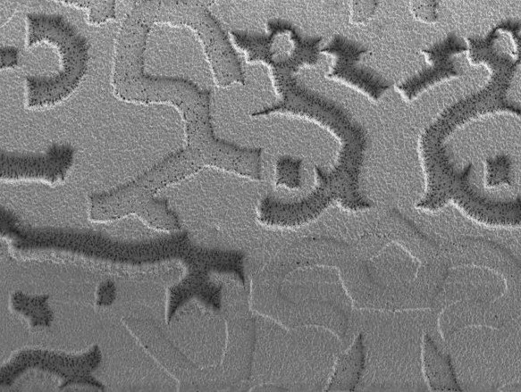
Engineered to influence a biological response at every level
-
Macro Surface
Mimics trabecular bone structure
-
Micro Surface
Provides pits for increased cellular attachment
-
Nano Surface
Elicits an endogenous cellular response

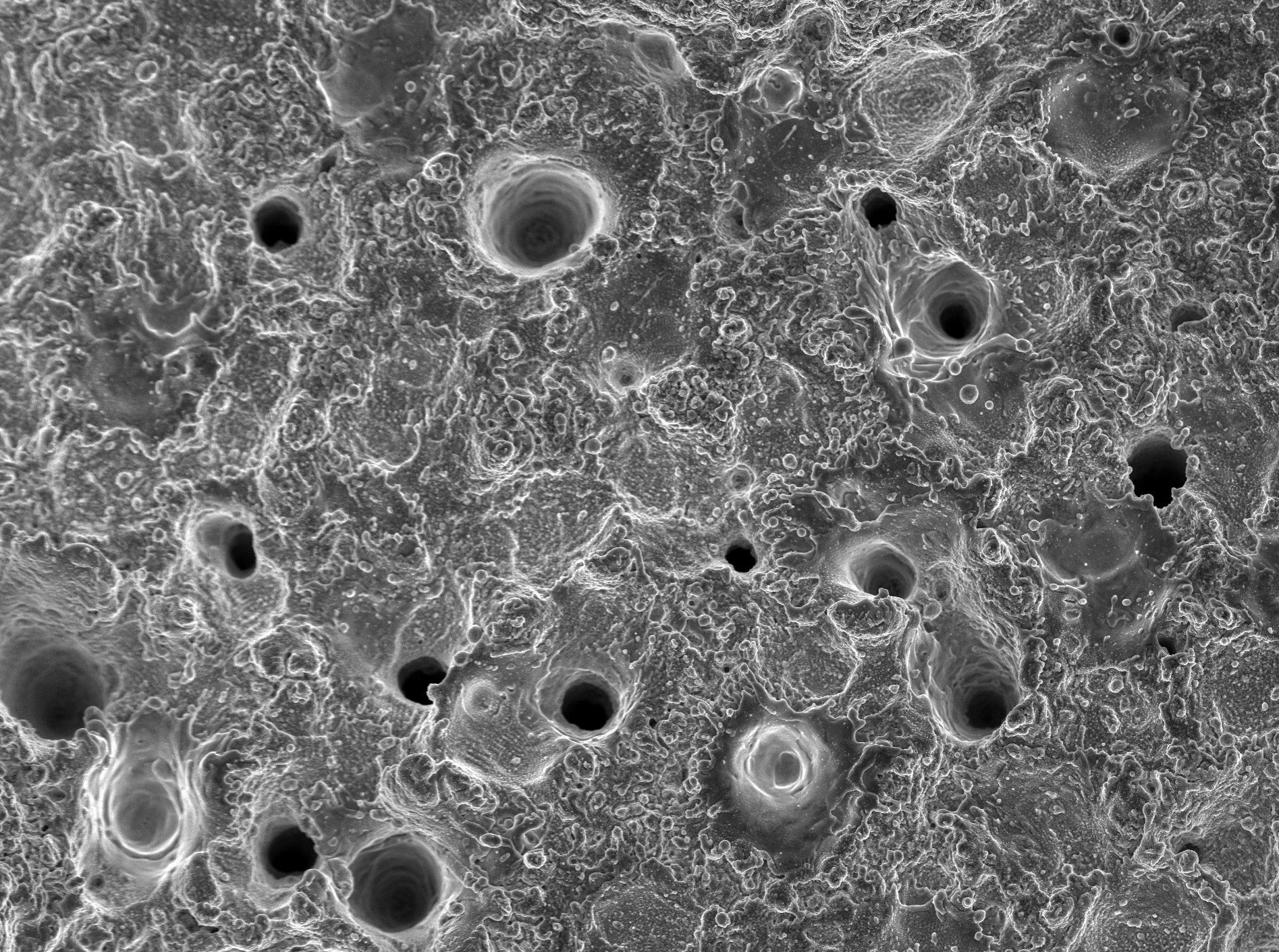
Pre Clincial Data for BioBraille™
The BioBraille™ surface demonstrated increased, rapid cell attachment when compared to smooth titanium.
Cellular Attachment | Day 14 | ~10x Magnification
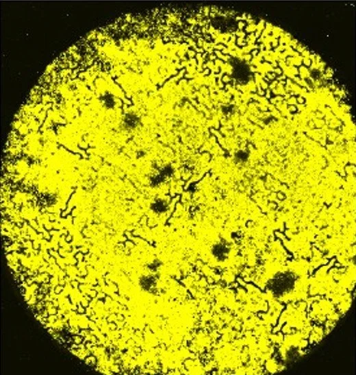
BioBraille™
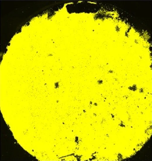
Smooth Titanium
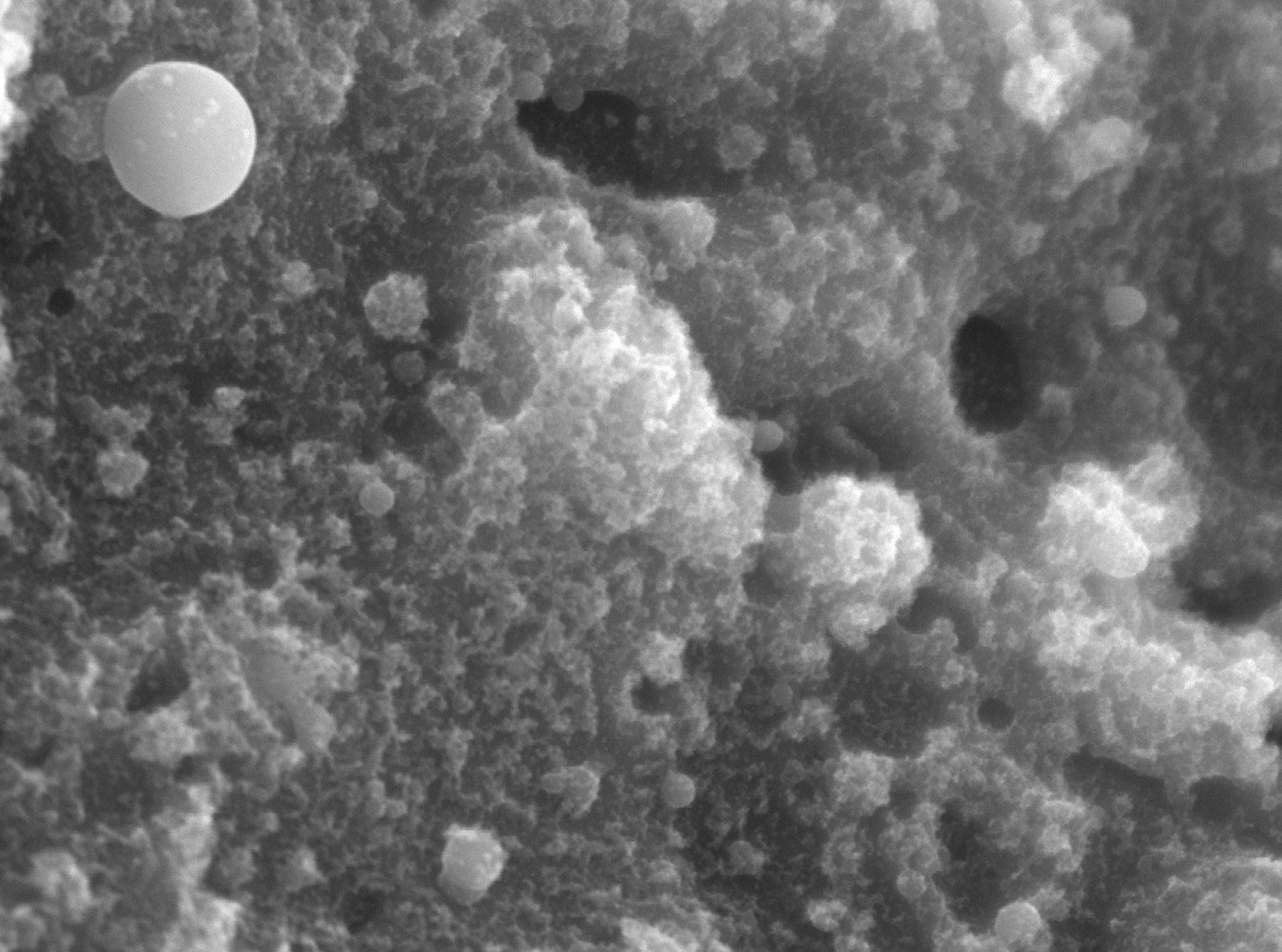
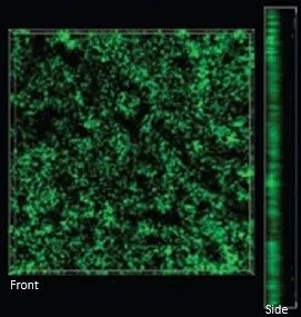
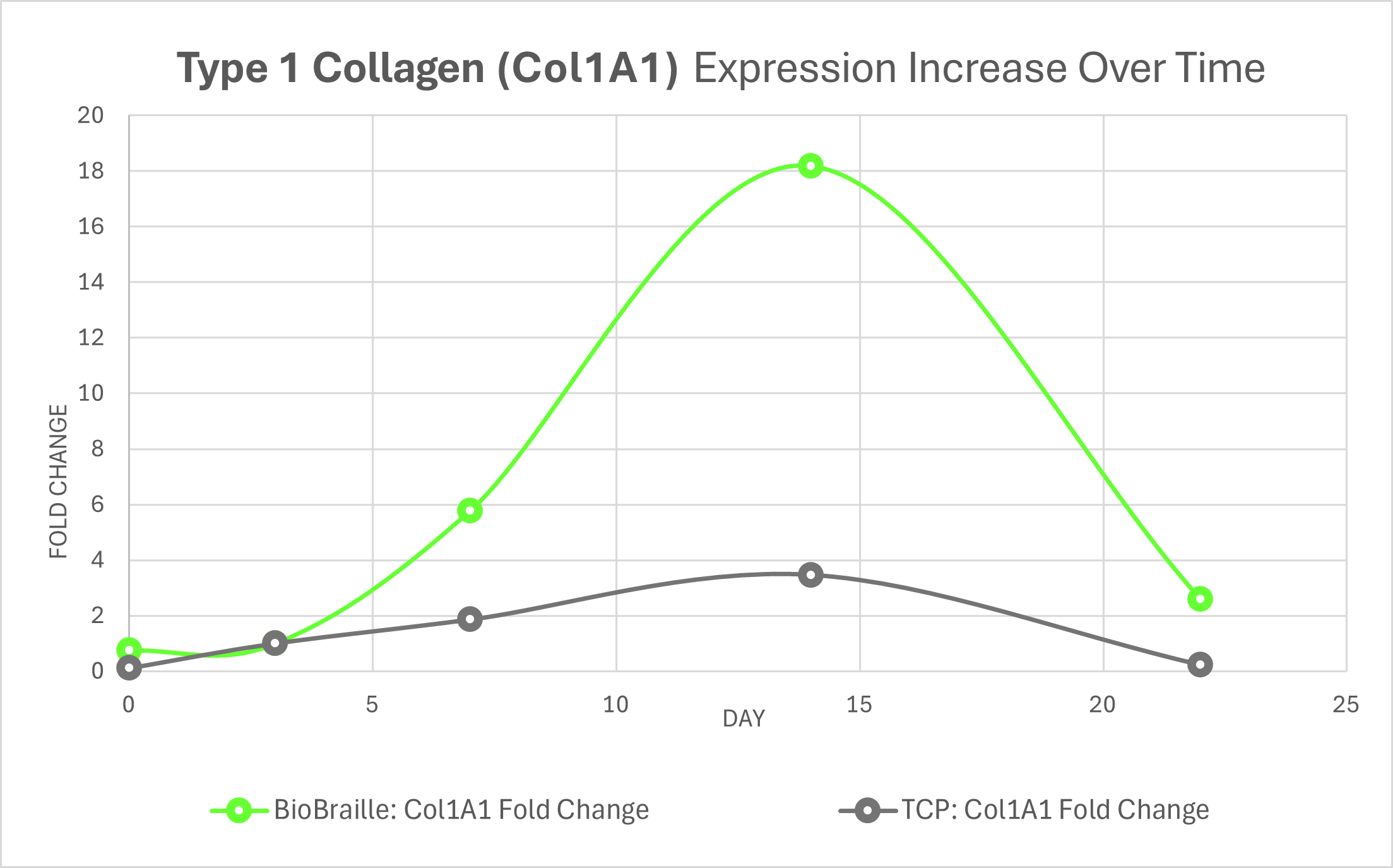
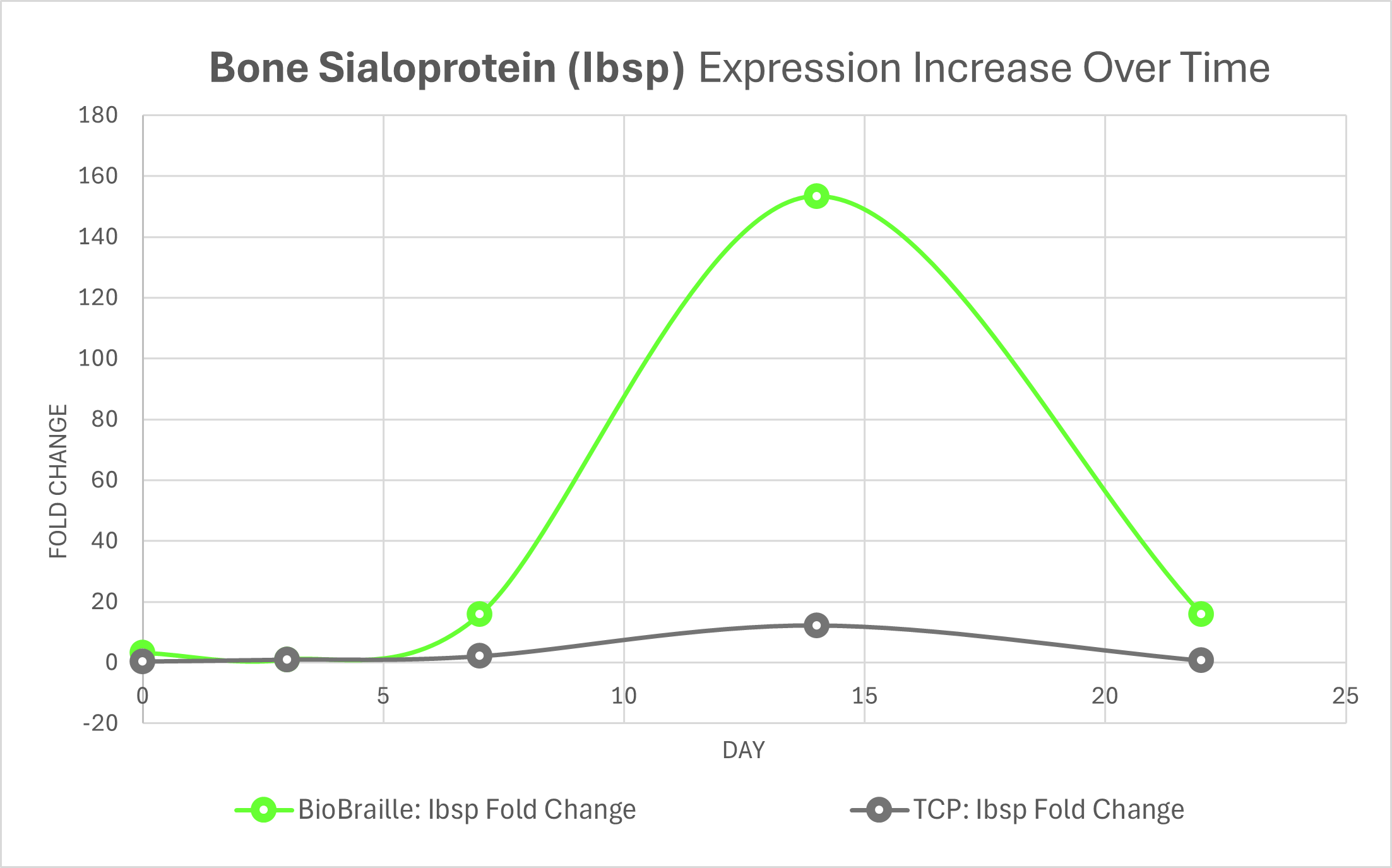
The BioBraille™ surface has also demonstrated increased expression of Type 1 Collagen, an early marker that is essential for bone formation as the dominant structural protein of bone extracellular matrix (ECM), as well as increased expression of Bone Sialoprotein and Dentin Matrix Protein, well-established markers of osteogenic differentiation and active bone matrix formation.
4D Projection of Cellular Expression | Day 14
BioBraille™ Surface (Front) | Depth of Proliferation (Side)
Bone Sialoprotein
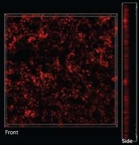
Dentin Matrix Protein
Our Femtosecond Laser
Femtosecond laser processing is an advancement in technology that allows Spectrum to “program” a macro, micro, and nano surface on selected areas.
Subtractive
Dimensionally accurate
Highly reproducible
Future Innovation Opportunities
Spectrum has enhanced the precision and versatility of nanotechnology integration by enabling targeted application of its proprietary BioBraille™ surface. This approach allows for strategic placement on implant areas requiring immediate bone contact and fusion, while preserving smooth, polished, or articulating surfaces to maintain optimal mobility and performance.
“BioBraille™ represents a transformative evolution in nanotechnology, with broad implications across the orthopedic landscape, including reconstruction, sports medicine, trauma, and dental applications. By combining scientific innovation with clinical insight, Spectrum is helping to redefine standards of care.” - Kelly Shelton, CEO.
Industry titans started it. We refined it – Nano, precisely placed.
*All images and technologies shown above are for illustrative purposes only. No device shown is claiming FDA approval or is available for purchase.